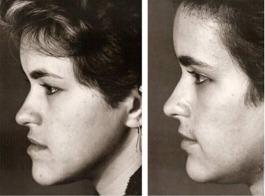
Ramus in human is having two surfaces and four borders. It has got two functions with it. The lateral surface of the ramus is flat, and the medial surface of it is oblique, providing the space of the nerves to enter the ridge. Margins are generally irregular, and a sharp spine surrounds the ridge. There is a rough surface inside, which usually stays behind the groove. The lower side of the border is thick and parallel to the bone. There is one posterior border too. It is supported by the deep concave shape. There is one coronoid process and one condyloid process involved in the Vertical Ramus Mandibular. The treatment is usually followed here when there is a fracture in the ramus.

Know the symptoms
The damage is usually known as jaw fracture, where the mandibular bone is damaged or broken inside. The first thing that the doctors or the surgeon will do here is the identification of the fracture spot. It is generally found in the Vertical Ramus Mandibular. In medical terms, they are stated to be Alveolar and Condylar. It is really painful and is caused due to some accidents. The common symptoms to understand the fracture will help you to understand when to go for a doctor.
-
Pain in the jaws and unable to chew food is the first of all the symptoms. In fact, the teeth here will be dislocated and hence you will have to be extra careful in the case.
-
Numbness is the next thing that you will feel. The damage or the fracture is going to yield pressure on the nerves, resulting in the numbness. You will also find a swollen nature in the facial muscles.
-
Bleeding from ear canals is also not astonishing. Since the Vertical Ramus Mandibular hits the eardrums, bleeding and even damage of the listing aid is not impossible.
Treatment Style
The treatment that you need to pursue starts with imaging. Radiography of the teeth is the first thing that you will have to point out. It can be added with Panoramic radiography or Computed Tomography. Depending on the complication that you are having. It depends on the doctors understanding of the shapes of the bones and the obligation it is facing from the bones. Treatment starts after the radiography, where the patient might feel jeopardized by the effect. Treatment has three steps involved. The first one is the force application to set the bones. The second is a reduction, where the bone edges are approximated. It can be done with an open or closed technique. Finally, the fixation is involved. It can be fixed internally and externally too.
The timing constraint in the treatment is the biggest challenge. A days delay in the diagnosis and surgery can damage the structure of the bones forever. So, if you find the above-stated symptoms then immediately consult a dentist, who will take care of the things and will recommend the immediate tests, following which, he will start the treatment. So, do not leave these things for a day even, a days gap in treatment can be dangerous.


